Most people expect an eye test to involve reading letters from a chart. In reality, that is only one part of a comprehensive eye examination.
Your optometrist runs several tests to check your vision, look at the health of your eyes, and spot any issues before they start affecting your sight.
This guide walks you through the key tests in a comprehensive eye exam, so nothing about your appointment comes as a surprise.
Quick Overview
A modern sight test brings together a few key tools to map out your vision and catch any issues early. Here is what it usually includes:
- Visual Acuity: Measuring clarity at specific distances.
- Refraction: Determining exact lens prescriptions.
- Tonometry: Testing eye pressure.
- Visual Field Mapping: Checking peripheral vision.
- Retinal Imaging: Capturing detailed images of the back of the eye.
- OCT Scanning: Analysing deep-tissue retinal layers.
- Digital Microscopic Examination: Inspecting the front surface of the eyes
What Is an Eye Test and Why Is It Important?
An eye test is a full medical evaluation of your eye health. It does more than just see how well you can read. It looks for early warning signs of systemic diseases and conditions like:
- Macular degeneration
- Diabetic retinopathy
- Glaucoma
- Cataracts
These serious issues usually develop quietly. You might not feel any pain or see any changes until permanent damage occurs. Regular check-ups help protect your sight before a minor issue turns into a permanent problem.
What Does an Eye Test Involve?
Your eye test starts with a simple chat in which optometrists discuss your:
- Health history
- Family background
- Daily habits.
Once they understand your eyes, your health, and how you use them day-to-day, they move on to the technical side of the exam to see how your eyes are actually performing.
After establishing a clear picture of your life, they move into the technical side of the exam to see how your eyes are actually performing.
7 Essential Tests Your Optometrist Uses During an Eye Exam:
Here are the tests optometrists rely on to get a clear picture of your eye health. The results of these tests guide the next steps, whether you just need a fresh pair of glasses or a specific plan to manage a newly discovered issue:
| Test | What It Checks |
|---|---|
| Visual Acuity | Clarity of vision at various distances |
| Refraction | Precise lens prescription requirements |
| Tonometry (Eye Pressure Test) | Signs of ocular hypertension or glaucoma |
| Visual Field | Peripheral awareness and neurological pathways |
| Retinal Imaging | Detailed map of the retina and blood vessels |
| OCT Scan | Cross-sectional thickness of retinal layers |
| Digital Microscope | Health of the cornea, iris, and crystalline lens |
Visual Acuity Test
This test figures out the smallest letters you can read from 6 metres away. The result is written as a fraction, like 6/6. If you score 6/12, it means you have to be 6 metres away to read what someone with standard vision sees from 12 metres.
In our clinic, patients often feel their vision is fine but don’t realise they have been subconsciously squinting or straining to compensate for minor blur. Visual acuity tests identify such focusing issues and help patients get the right glasses to clear their vision.
Even if your eyes seem healthy, it is worth having this test every two years so you are not unknowingly living with eye strain.
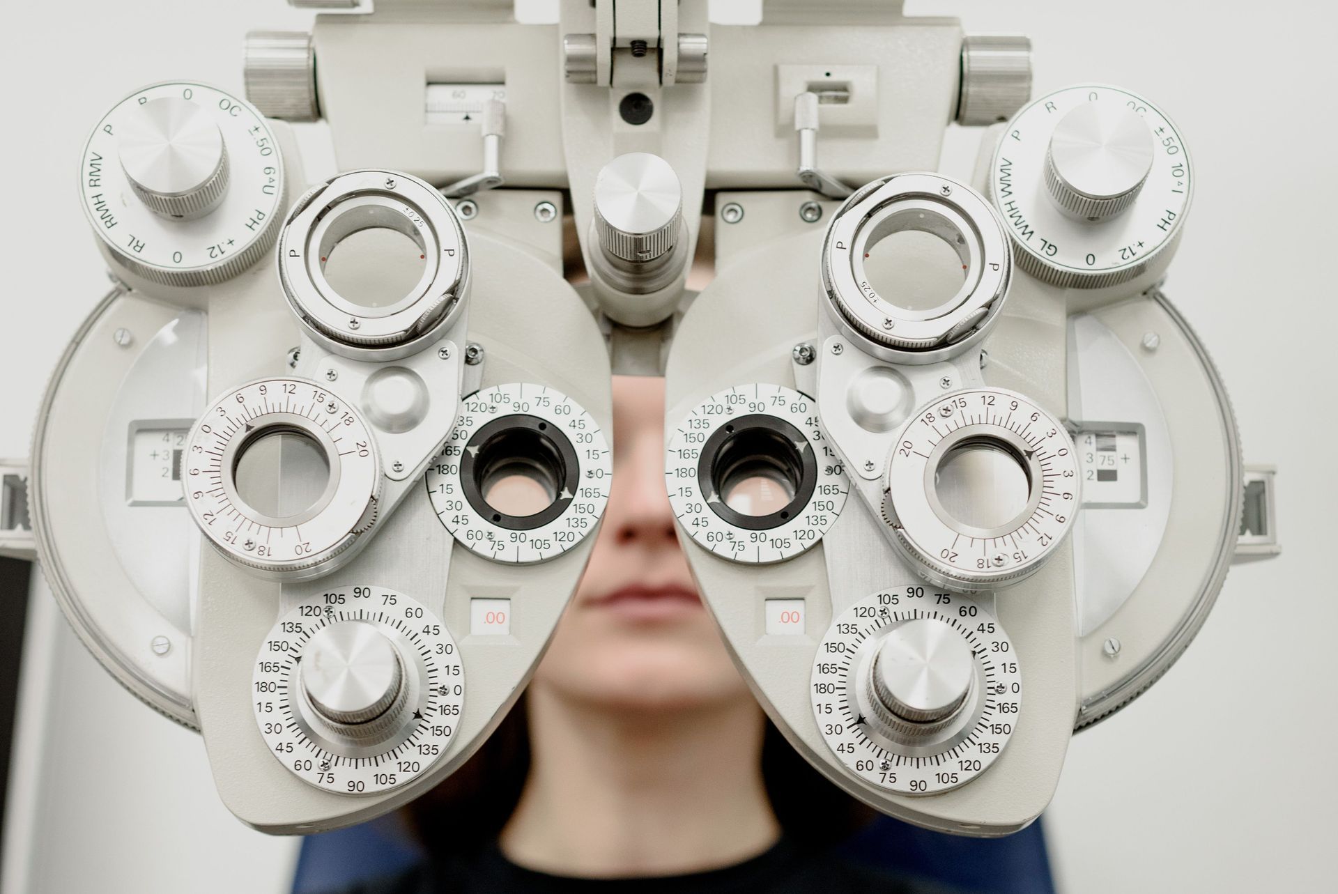
Refraction Assessment
This test finds your perfect prescription. By trying different lenses with a phoropter, optometrists map out exactly how your eyes focus.
Many people just settle for glasses from retail shops, but when your astigmatism is corrected by even a few degrees, the difference is huge. It is common for patients to be surprised at how much sharper the world looks once their vision is properly dialled in with a refraction test.
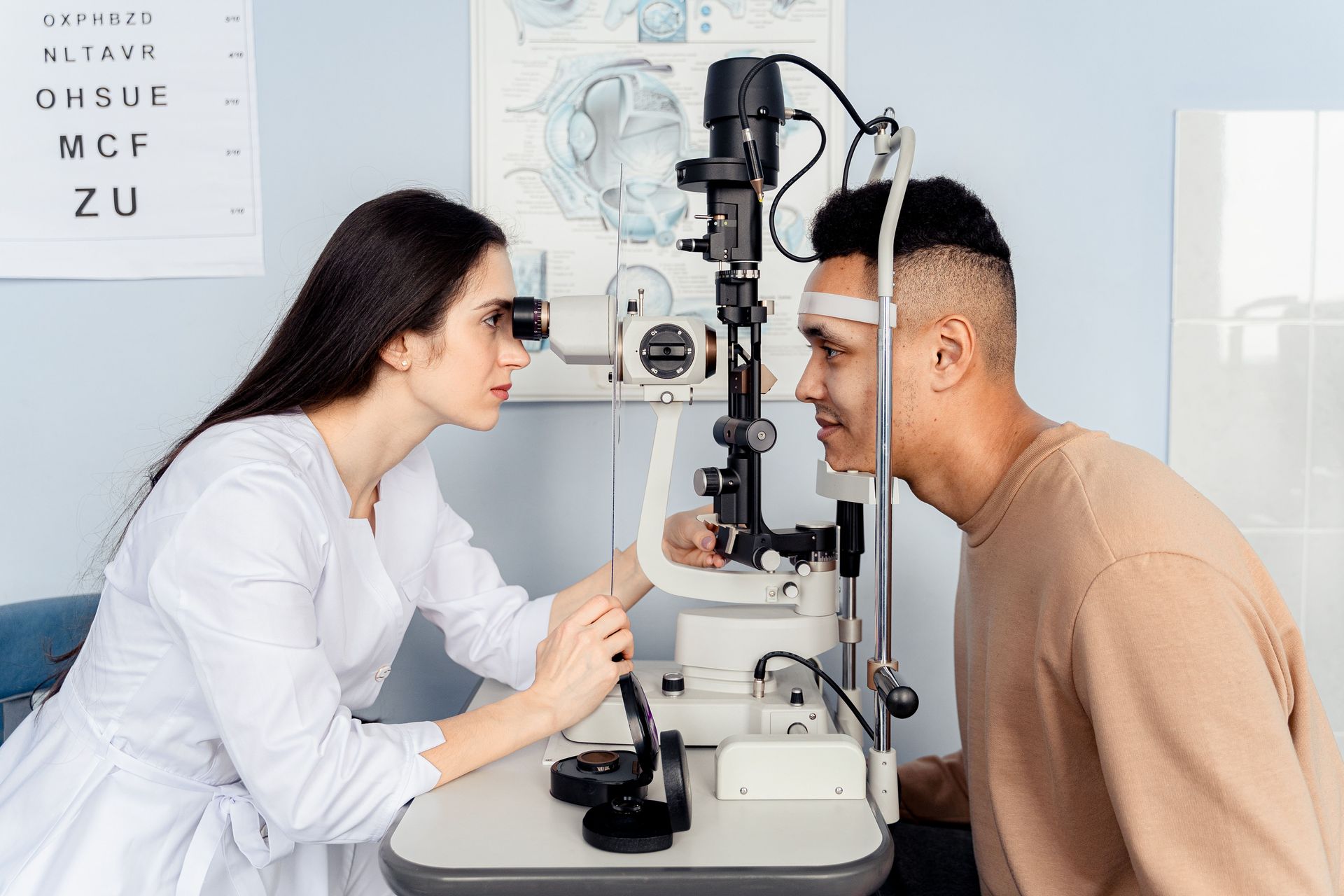
Tonometry (Eye Pressure Test)
In this test, optometrists use a tonometer to check the fluid pressure inside your eyeball. Normal intraocular pressure typically ranges between 10 and 21 mmHg, although some people develop glaucoma with pressures within this range and others have higher pressures without eye damage. If the number creeps higher, it might mean fluid isn't draining the way it should.
A patient might feel absolutely no discomfort but still have an elevated pressure of 25 mmHg. The test clarifies the pressure issues early on, and the medical provider may prescribe simple preventative drops. These drops keep the optic nerve healthy and help avoid the need for more invasive treatments later on.
Visual Field Testing
This test maps out your entire range of sight while you stare at one spot. It helps optometrists find blind spots, known as scotomas, in your side vision. These spots may indicate nerve damage.
We once had a patient who only noticed a slight issue while driving, but this test quickly identified a clear blind spot in their peripheral vision, and we referred them to a specialist right away.
Retinal Imaging
The procedure is like a high-tech photo of the back of your eye. Retinal imaging captures a detailed, ultra-wide photograph of the back of your eye. At our clinic, the Zeiss Clarus ultra-wide camera captures far more of the peripheral retina than a standard image, which helps detect disease earlier and keeps a permanent record to track changes over time.
Instead of just looking in with a light, optometrists take a wide-field picture that stays in your file. This test allows them to spot immediate issues and track tiny changes in your blood vessels over time.
For instance, in patients with high blood pressure, we often see narrowed or strained retinal arteries, which allows us to suggest a quick check-up with their primary care doctor.
OCT Scanning
Optical Coherence Tomography, or OCT, uses light waves to give optometrists a cross-section of your retina. While a standard retinal image captures the surface view of the retina at the back of the eye, an OCT is often called an "optical biopsy" because it looks deeper and lets the optometrist see hidden tissue layers that a photo cannot show.
In our experience, this scanning has saved central vision for many people by catching fluid buildup early, long before it would show up on a standard exam.
Slit Lamp Examination (Digital Microscope Test)
The result is a high-magnification 3D view of the front of your eye. Using a slit lamp microscope, the optometrist examines the cornea, conjunctiva, eyelids, and tear film to check for dryness, inflammation, and early surface damage.
This test explains why your eyes feel gritty, watery, or tired, and it guides the right treatment. If the exam points to dry eye, your optometrist can talk you through a management plan suited to your eyes, which may include:
- Specific eyelid cleaning
- Warm compresses
- More omega-3 fatty acids in your diet
- Regular screen breaks to encourage blinking
The right plan depends on what the exam finds, so it is best confirmed with your optometrist rather than self-managed.
Comprehensive Eye Exam vs Standard Vision Check: What's the Difference?
Many patients confuse a basic vision check with a full medical evaluation, but the two services serve completely different purposes. A vision check is usually just to get you a new pair of specs. A comprehensive eye exam is a deeper medical look at the structure and health of your eye tissues.
Here is exactly what you get with each option:
| Feature | Standard Vision Check | Comprehensive Eye Exam |
|---|---|---|
| Primary Goal | Glasses prescription | Disease detection & health monitoring |
| Diagnostic Scope | Basic sight chart | Digital imaging, OCT, & pressure tests |
| Health Focus | Refractive error (blurriness) | Glaucoma, retinal, & corneal health |
| Outcome | New spectacles | Full eye health record & diagnosis |
When Should You Get Your Eyes Checked?
If you notice any of the following signs, don't wait for your next check-up and consult an optometrist right away:
- Suddenly blurred or warped vision.
- Frequent flashes of light or "floaters" in your view.
- Ongoing eye pain or a feeling of pressure.
- Constant headaches after you finish working at your computer.
If everything seems fine, just stick to a routine two-year checkup. It is the best way to make sure nothing changes without you noticing.
Conclusion
Think of a full eye exam as a proper health check, not just a way to update your glasses. Reading the chart is part of it, but the real value is in the scans that catch quiet problems long before you would feel them. So keep your tests regular, and do not wait for something to go wrong.
If you would like that kind of thorough checkup for your eyes, our family-run team at Nazarian Optometrists has been caring for Western Sydney since 1985. Book an appointment today, and let us give your vision the care it deserves.
Frequently Asked Questions
Should I Get an Eye Test Even If My Vision Seems Fine?
Yes. Many eye diseases are silent. Regular testing is the only way to detect conditions like glaucoma before they cause irreversible vision loss.
Are Eye Tests Bulk Billed in Australia?
Comprehensive eye exams are generally bulk-billed for eligible Medicare cardholders, making medical-grade eye care accessible to the community.
Is an Eye Test Painful or Uncomfortable?
Not at all. Most diagnostic tests are non-contact. You might feel a tiny puff of air during the pressure test, but it is quick and completely harmless.
How Long Does a Full Eye Test Take?
A comprehensive exam usually takes around 30 to 45 minutes, depending on which diagnostic scans you need. We will always talk you through each step as we go.
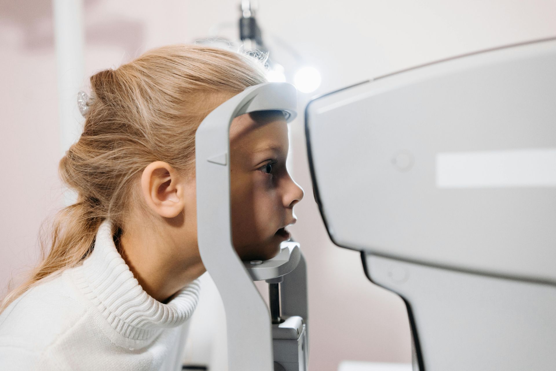
How Often Should Children Have Their Eyes Tested?
Most children benefit from a check before they start school, then every one to two years, or sooner if you notice squinting or sitting very close to screens. Children's exams are bulk billed for eligible Medicare patients.
Can an Eye Test Detect Other Health Conditions?
Yes. Retinal imaging can reveal early signs of diabetes and high blood pressure through subtle changes in the eye's blood vessels, which is one reason regular exams matter beyond your prescription.