Have you recently noticed a brown, black, grey, or blue spot on the white part of your eye? If so, you might be worried about why it appeared and how to get rid of it. It’s natural to worry, especially when you see it for the first time in your life.
Many dark spots are eye freckles or age-related changes. However, a new or changing mark may need closer examination. So, let’s learn what a black spot on the eye white looks like, what may be the cause behind it, and whether it affects vision.
We will also walk you through possible treatment options for dark spots on eye whites so you know how to recover from them. Let’s get started.
What Is a Dark Spot on Eye White?
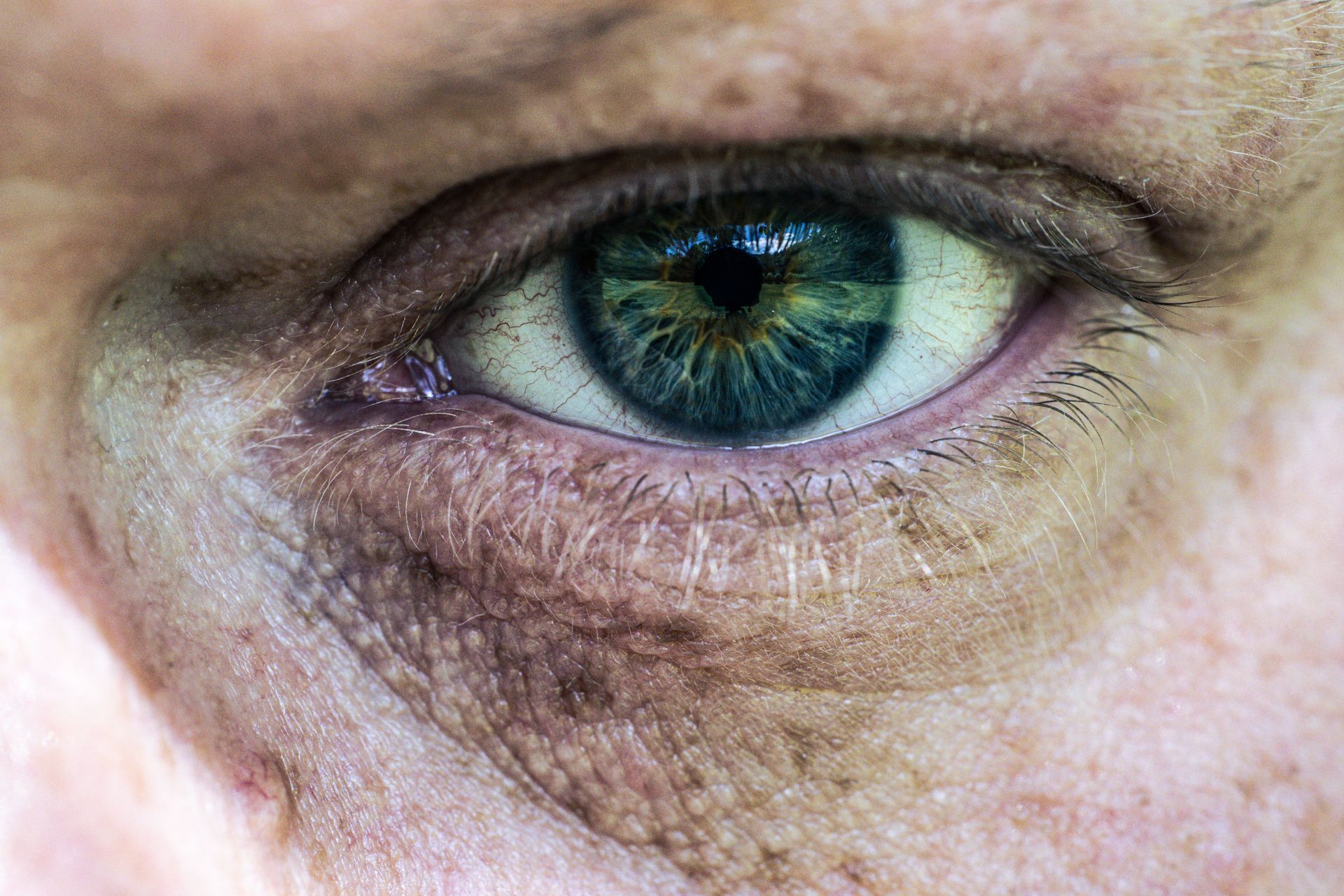
A dark spot on the white of the eye may be due to pigmentation, bleeding, trapped material, or a tissue change. It commonly appears on the conjunctiva, which is the thin, clear membrane covering the sclera, or white part of your eye.
Some spots develop during childhood but only become noticeable years later, in photographs or in a mirror. Others develop later because of sun exposure, ageing, inflammation, injury, or a growth on the eye’s surface.
We often see patients who arrive worried that any brown or black mark on the eye must be cancerous. In most cases, our examination shows a harmless eye freckle, an age-related change, or some other benign condition.
How to Recognise Different Types of Dark Spots
Dark spots can vary in colour, shape, thickness, and location. These features may provide clues, but only a clinical eye examination can establish the likely cause. Here are different types of dark spots:
Brown Spots
Brown spots are often linked to natural pigmentation or a conjunctival nevus, commonly called an eye freckle.
A conjunctival nevus may appear:
- Light or dark brown
- Round or oval
- Flat or slightly raised
- Clearly outlined
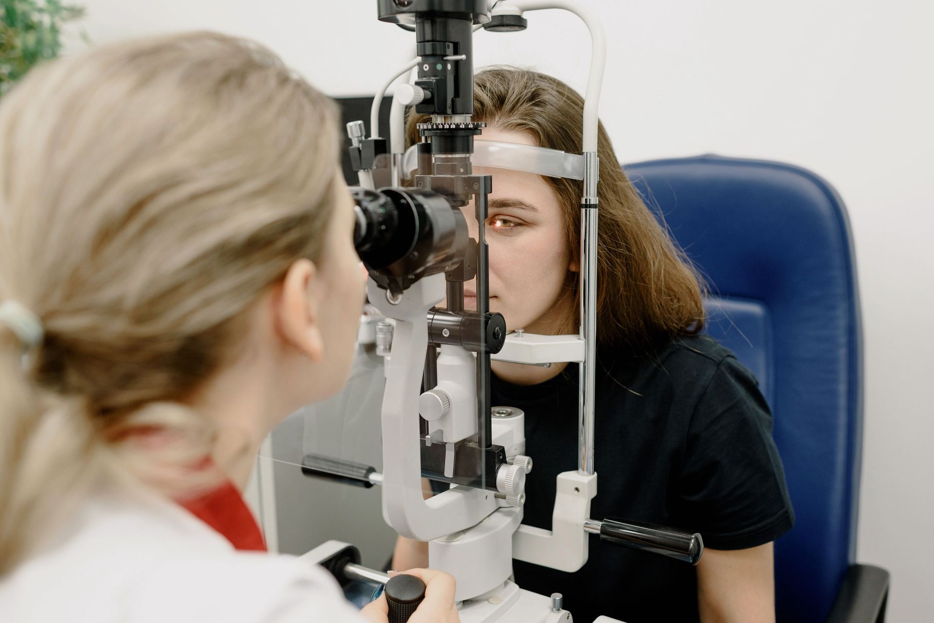
Some nevi contain tiny clear cysts that an optometrist can see through a slit-lamp microscope. These cysts often suggest that the lesion is benign, although the entire spot still requires clinical assessment. Conjunctival nevi are the most common melanocytic tumours of the conjunctiva.
Black Spots
A black spot on the white of the eye may be a deeply pigmented nevus, embedded debris from an earlier injury, or another pigmented lesion.
It needs closer attention if it:
- Becomes darker
- Grows quickly
- Develops uneven edges
- Starts bleeding
- Shows new blood vessels
Colour alone does not reveal whether a spot is dangerous. A stable black mark may be harmless, while a lighter lesion that changes rapidly may need referral.
Grey Spots
Grey spots can occur with age-related thinning or degeneration of the sclera. Senile scleral plaques, for example, often appear as grey areas near the points where the horizontal eye muscles attach.
An old metallic foreign body can also leave grey or dark pigmentation. Your optometrist may ask about past work with grinding tools, machinery, metal, or building materials.
Blue or Slate-Coloured Spots
A bluish tint often means the pigment lies deeper within the sclera itself rather than in the surface conjunctiva. Senile scleral plaques, which are more common after age 60, typically have a bluish-grey appearance.
What Causes Dark Spots on the Whites of the Eyes?
Dark spots on the whites of the eyes have several possible causes. Many are harmless, but some need monitoring or specialist treatment. Let’s walk through the potential causes of dark spots.
Conjunctival Nevus (Eye Freckle)
A conjunctival nevus is a benign collection of pigment-producing cells called melanocytes. It often appears during childhood or early adulthood and may remain unchanged for decades.
In our clinic, patients often tell us they have only just noticed a brown spot even though it may have been present for years. A family member may point it out, or the patient may see it clearly in a close-up photograph for the first time.
Pinguecula
A pinguecula is a small, raised area that usually develops beside the cornea. It often looks cream, yellow, or light brown rather than truly black.
Long-term exposure to sunlight, wind, dust, and dry conditions may contribute to its development. It may become red or uncomfortable when inflamed, but it does not grow across the cornea.
Pterygium
A pterygium is a fleshy, triangular growth that starts on the conjunctiva and extends towards the cornea. It may contain visible blood vessels and areas of brown pigmentation.
Unlike a pinguecula, a pterygium can grow onto the cornea. A larger growth may change the shape of the cornea, cause astigmatism, or obstruct vision.
Subconjunctival Haemorrhage
According to the Mayo Clinic, subconjunctival haemorrhage occurs when a small blood vessel breaks beneath the conjunctiva. It usually starts as a bright red patch. During healing, it may turn dark red, brown, yellow, or almost black.
These haemorrhages often look alarming but generally clear without treatment. Repeated episodes should still be discussed with a health professional, particularly if you take blood-thinning medication or have high blood pressure.
Primary Acquired Melanosis (PAM)
Primary acquired melanosis, or PAM, usually appears as a flat brown area that develops during adulthood. It often affects one eye and may have poorly defined or irregular edges.
PAM without atypical cells may be benign. PAM with atypia can be precancerous and requires specialist assessment. A visual examination cannot confirm whether atypia is present because that distinction depends on tissue analysis.
Conjunctival or Ocular Melanoma
Conjunctival melanoma is a rare but serious malignant tumour. The American Academy of Ophthalmology describes this disease as a potentially life-threatening condition because it can spread to other parts of the body if left untreated. It may develop from PAM, an existing nevus, or without a previous visible lesion.
Features that raise concern include:
- Rapid growth
- Increasing thickness
- Irregular pigmentation
- Prominent blood vessels
- Bleeding
- Fixation to deeper tissue
These signs do not prove that a lesion is melanoma, but they warrant prompt referral to an ophthalmologist.
Foreign Body Pigmentation / Debris
Another common cause of black spots on the white part of the eye is external debris. Fragments of metal, graphite, soil, stone, or other material can become trapped in or beneath the conjunctiva after an injury. A retained particle may look like a tiny black dot.
Even when pain settles, an eye injury involving metalwork or high-speed debris needs prompt assessment because a fragment may have entered deeper structures.
Medication-Related Pigmentation
Some medicines can cause changes in conjunctival or scleral colour after long-term use. Some examples of such medicines include Minocycline and Amiodarone.
If you see a dark spot due to a medication, do not stop the prescribed medicine because of eye pigmentation. Instead, ask your optometrist and prescribing doctor to review the change and decide whether further testing is appropriate.
Senile Scleral Plaques
Senile scleral plaques are age-related grey areas that develop on the white of the eye, usually near the horizontal eye muscles. They are generally harmless and do not require treatment. However, an examination can confirm that the mark matches this common age-related pattern.
Congenital Ocular Melanocytosis
Congenital ocular melanocytosis causes blue-grey pigmentation within deeper eye tissues and is usually present from birth. Because this condition can affect more than the visible surface, an optometrist may recommend regular dilated eye examinations rather than only photographing the external mark.
Symptoms That May Occur Alongside a Dark Spot
Many dark spots cause no discomfort. Symptoms usually arise when the surrounding eye surface becomes dry or inflamed, or when another eye condition is present.
Possible symptoms include:
- Redness or irritation
- Dryness
- A gritty or foreign-body sensation
- Excessive watering
- Light sensitivity
- Pain
- Blurred vision
- Changes in the spot’s colour or size
- Bleeding from the lesion
A harmless conjunctival nevus should not normally cause severe pain or sudden vision loss. Symptoms of that kind require assessment for another underlying problem.
When Is a Dark Spot an Eye Emergency?
Most pigmented spots are not emergencies. However, a sudden or changing mark needs prompt attention, especially when it occurs with pain, bleeding, injury, or altered vision.
A painless lesion can still need investigation if it grows, changes colour, or develops irregular edges. Pain is only one warning sign and should not be used as the sole measure of seriousness.
Seek urgent eye care if the spot:
- Appears suddenly
- Enlarges over weeks or months
- Develops irregular borders
- Contains several colours
- Bleeds without an obvious injury
- Appears after metalwork or eye trauma
- Occurs with severe pain
- Develops alongside sudden vision loss
Do Dark Spots on Whites of Eyes Affect Vision?

Most dark spots on the white of the eye do not affect vision because they sit on the conjunctiva rather than the clear cornea or the structures that process sight. However, in some cases, they can cause vision problems, although this is less common than many people believe. In fact, the dark spot is often mistaken for the main cause, when in reality it is usually other underlying eye conditions that are responsible.
A dark spot may be one of the potential causes of blurry vision when:
- A pterygium grows onto the cornea.
- Surface irritation disrupts the tear film.
- A lesion extends into deeper tissue.
- Another eye condition occurs at the same time.
Many people assume the visible spot must be causing their blurred vision. However, a thorough eye examination often shows that the mark is stable and harmless. Instead, dry eye, a changing glasses prescription, or an early cataract may be responsible for the reduced clarity.
For that reason, the spot should not be assessed in isolation. A comprehensive eye examination includes checking visual acuity, your prescription, the eye surface, and the internal eye structures to identify the true cause of any vision changes.
How Are Dark Spots on the White of the Eye Treated?
Treatment depends entirely on what the spot actually is, and most cases need far less intervention than people expect. According to
Cleveland Clinic and
LivHospital, here are the possible treatments that patients might have to undergo depending on the type of dark spot on the white part of their eye:
| Condition | Typical First-Line Treatment | Surgery Needed? |
|---|---|---|
| Conjunctival nevus | Observation, periodic photography | Rarely, unless suspicious changes appear |
| Pinguecula | Lubricating drops, sometimes short-term steroid drops | Rarely |
| Pterygium | Lubricating drops; surgery if vision affected | Often, if growth crosses onto the cornea |
| Subconjunctival haemorrhage | None; monitor for recurrence. | No |
| PAM | Regular monitoring; biopsy if atypia suspected | Sometimes |
| Conjunctival melanoma | Surgical removal, follow-up oncology care | Yes. |
- Observation and Monitoring: For a stable nevus or a low-risk pinguecula, the standard approach is simply watching it over time with periodic photographs to catch any change early.
- Lubricating Eye Drops for Dryness-Related Irritation: When a spot brings mild grittiness or dryness, preservative-free lubricating drops usually settle the irritation within days.
- Treating Pinguecula and Pterygium: Beyond lubricants, short courses of anti-inflammatory drops can calm redness and swelling during flare-ups, particularly after windy or dusty conditions.
- Managing Subconjunctival Haemorrhage: No active treatment is generally needed, since the blood reabsorbs naturally. A doctor may check blood pressure or clotting factors if it happens repeatedly.
- Surgical Removal: A pterygium that grows onto the cornea and distorts vision, or a nevus that shows worrying changes, may be surgically removed, often as a same-day outpatient procedure.
- Treatment for Suspected Melanoma: If melanoma is suspected or confirmed, treatment moves quickly to surgical excision, often paired with additional therapies and long-term specialist follow-up to watch for recurrence.
Wrapping It Up
Most dark spots on the whites of the eyes are harmless, especially when they have remained stable for years and cause no symptoms. Still, you should not rely on colour or online photographs to identify the cause. Benign freckles, old bleeding, foreign material, acquired pigmentation, and more serious lesions can look similar without magnification.
So, you should monitor any new lesion carefully, especially if it changes in size, shape, or colour over time. At Nazarian Optometrists, our experienced team has assessed eye surface lesions for more than 30 years. Using advanced imaging technology, including OCT scanning and ultra-wide retinal photography, we can determine whether a dark spot is harmless or requires specialist referral.
Book a comprehensive eye examination if you have noticed a new or changing dark spot and want a clear, personalised assessment.
Frequently Asked Questions
Can a black spot on the white of my eye go away on its own?
A dark mark caused by a subconjunctival haemorrhage may fade as the trapped blood clears. A conjunctival nevus, congenital pigmentation, or embedded foreign material usually remains unless treated.
Is a brown spot on the white of my eye cancer?
Most brown spots are not cancerous. However, a new lesion or one that changes in size, shape, thickness, or colour requires examination because appearance alone cannot rule out a serious cause.
What causes a sudden dark spot to appear in the eye?
A suddenly visible mark may come from bleeding beneath the conjunctiva, eye trauma, trapped debris, or a lesion that has recently changed. Prompt assessment is advisable when the cause is unclear.
Are dark spots on the eye more common with age?
Some types become more common with age, including senile scleral plaques and primary acquired melanosis. Conjunctival nevi often appear much earlier in life.
Is it safe to wear contact lenses if I have a dark spot?
Contact lens wear depends on the cause and whether the surface is inflamed. Stop wearing the lenses and arrange an assessment if the eye feels painful, red, gritty, or unusually sensitive to light.